A Review of Current Cell Annotation Systems for Histopathology Images

Authors

  • Chai Ling Teoh Biomedical and Bioinformatics Engineering (BBE) Research Group, Centre for Multimodal Signal Processing (CMSP), Tunku Abdul Rahman University of Management and Technology (TAR UMT), Setapak, 53300 Kuala Lumpur, Malaysia
  • Xiao Jian Tan Biomedical and Bioinformatics Engineering (BBE) Research Group, Centre for Multimodal Signal Processing (CMSP), Tunku Abdul Rahman University of Management and Technology (TAR UMT), Setapak, 53300 Kuala Lumpur, Malaysia
  • Khairul Shakir Ab Rahman Department of Pathology, Hospital Tuanku Fauziah, 01000 Kangar, Perlis, Malaysia
  • Ikmal Hisyam Bakrin Department of Pathology, Faculty of Medicine and Health Sciences, Universiti Putra Malaysia, UPM Serdang, 43400 Serdang, Selangor, Malaysia
  • Kam Meng Goh Department of Electrical and Electronics Engineering, Faculty of Engineering and Technology, Tunku Abdul Rahman University of Management and Technology (TAR UMT), Setapak, 53300 Kuala Lumpur, Malaysia
  • Wan Zuki Azman Wan Muhamad Institute of Engineering Mathematics, Universiti Malaysia Perlis (UniMAP), 02600 Arau, Perlis, Malaysia
  • Chee Chin Lim Sports Engineering Research Centre (SERC), Universiti Malaysia Perlis (UniMAP), 02600 Arau, Perlis, Malaysia
  • Wai Loon Cheor Biomedical and Bioinformatics Engineering (BBE) Research Group, Centre for Multimodal Signal Processing (CMSP), Tunku Abdul Rahman University of Management and Technology (TAR UMT), Setapak, 53300 Kuala Lumpur, Malaysia
  • Thakerng Wongsirichot Division of Computational Science, Faculty of Science, Prince of Songkhla University, Hat Yai, Songkhla 90110, Thailand

DOI:

https://doi.org/10.37934/ard.135.1.88100

Keywords:

Cell annotation, cell labelling, cell segmentation, nuclei segmentation, system, histopathology, digital pathology

Abstract

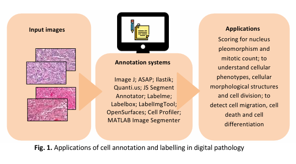
Histopathology techniques offer a unique way to study the structural and functional characteristics of biological model systems such as cultured cells, tissues and organoids. As the field of histopathology advances and more complex properties of living organisms are revealed through novel assays, there is a growing need for image analysis methods that are robust and easy to use. In many histopathology image analysis workflows, the first step is to segment cell nuclei, as they serve as the fundamental block for identifying individual cells in histopathology images. Such methods are crucial in a range of research studies, from counting cells and tracking moving populations to localizing proteins, classifying phenotypes and profiling treatments. Here, this review intended to provide an updated cell annotation system for histopathology images using the predetermined search strings and a set of inclusion criteria. Accordingly, 11 cell annotation systems (i.e., Image J, ASAP, Ilastik, Quanti.us, JS Segment Annotator, Labelme annotation tool, Labelbox, LabelImgTool, OpenSurfaces Segmentation UI, Cell Profiler and MATLAB Image Segmenter) was included. An in-depth discussion on the background of each included system was provided in this review alongside a brief comparison across different cell annotation systems.

Downloads

Download data is not yet available.

Author Biographies

Chai Ling Teoh, Biomedical and Bioinformatics Engineering (BBE) Research Group, Centre for Multimodal Signal Processing (CMSP), Tunku Abdul Rahman University of Management and Technology (TAR UMT), Setapak, 53300 Kuala Lumpur, Malaysia

charlene611ling@gmail.com

Xiao Jian Tan, Biomedical and Bioinformatics Engineering (BBE) Research Group, Centre for Multimodal Signal Processing (CMSP), Tunku Abdul Rahman University of Management and Technology (TAR UMT), Setapak, 53300 Kuala Lumpur, Malaysia

tanxj@tarc.edu.my

Khairul Shakir Ab Rahman, Department of Pathology, Hospital Tuanku Fauziah, 01000 Kangar, Perlis, Malaysia

ksyakir@gmail.com

Ikmal Hisyam Bakrin, Department of Pathology, Faculty of Medicine and Health Sciences, Universiti Putra Malaysia, UPM Serdang, 43400 Serdang, Selangor, Malaysia

ikmalhisyam@upm.edu.my

Kam Meng Goh, Department of Electrical and Electronics Engineering, Faculty of Engineering and Technology, Tunku Abdul Rahman University of Management and Technology (TAR UMT), Setapak, 53300 Kuala Lumpur, Malaysia

gohkm@tarc.edu.my

Wan Zuki Azman Wan Muhamad, Institute of Engineering Mathematics, Universiti Malaysia Perlis (UniMAP), 02600 Arau, Perlis, Malaysia

wanzuki@unimap.edu.my

Chee Chin Lim, Sports Engineering Research Centre (SERC), Universiti Malaysia Perlis (UniMAP), 02600 Arau, Perlis, Malaysia

cclim@unimap.edu.my

Wai Loon Cheor, Biomedical and Bioinformatics Engineering (BBE) Research Group, Centre for Multimodal Signal Processing (CMSP), Tunku Abdul Rahman University of Management and Technology (TAR UMT), Setapak, 53300 Kuala Lumpur, Malaysia

wlcheor@gmail.com

Thakerng Wongsirichot, Division of Computational Science, Faculty of Science, Prince of Songkhla University, Hat Yai, Songkhla 90110, Thailand

thakerng.w@psu.ac.th

Downloads

Published

2025-06-26

How to Cite

Chai, L. T., Xiao, J. T., Ab Rahman, K. S., Bakrin, I. H., Kam, M. G., Wan Muhamad, W. Z. A., Chee, C. L., Wai, L. C., & Wongsirichot, T. (2025). A Review of Current Cell Annotation Systems for Histopathology Images. Journal of Advanced Research Design, 135(1), 88–100. https://doi.org/10.37934/ard.135.1.88100
سرور مجازی ایران Decentralized Exchange

Issue

Section

Articles

Most read articles by the same author(s)

فروشگاه اینترنتی